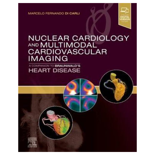
Title of the Book
▶ Click Image or Title to the Page on the Site
Nuclear Cardiology and Multimodal Cardiovascular Imaging,1/e
Product details
도서명: Nuclear Cardiology and Multimodal Cardiovascular Imaging,1/e
저 자: Marcelo Fernando Di Carli
출판사: Elsevier
ISBN : 9780323763035
출판일: 2022.01
판 형: Hardcover
판 수: 1/e
면 수: 464 page
Description
Recent years have seen numerous advances in cardiovascular nuclear medicine technology, leading to more precise diagnoses and treatment and an expanded understanding of the molecular basis for cardiac disease. Nuclear Cardiology and Multimodal Cardiovascular Imaging is a one-stop, comprehensive guide to the diagnostic and clinical implications of this complex and increasingly important technology. Part of the Braunwald family of renowned cardiology references, it provides cutting-edge coverage of multimodal cardiac imaging along with case vignettes and integrated teaching content—ideal for cardiologists, cardiology fellows, radiologists, and nuclear medicine physicians.
Key Features
- Features all the latest cardiovascular nuclear medicine studies with practical, evidence-based implications for personalized patient evaluation and treatment.
- Presents a consistent, patient-centered approach using integrated case vignettes correlated with specific nuclear medicine imaging findings. Discusses patient assessment criteria, risk factor criteria, pathology, evaluation criteria, outcomes, and other clinical implications.
- Covers a full range of imaging technologies, including SPECT/CT, PET/CT, and CT/MR hybrid radionuclide cardiovascular imaging studies.
- Addresses emerging clinical applications of nuclear imaging techniques for precision-based medicine, including targeted molecular imaging and cell therapies.
- Includes sections on instrumentation/principles of imaging; protocols and interpretation; applications in coronary artery disease, special populations, and heart failure; artificial intelligence, and more.
- Contains guidelines and appropriate use documents to provide appropriate context for clinicians.
- Features hundreds of high-quality figures including multimodal cardiac imaging studies, anatomic illustrations, and graphs.
- Provides Key Point summaries, 50 procedural videos, and 100 multiple-choice questions and answers to reinforce understanding and facilitate review.
- Enhanced eBook version included with purchase, which allows you to access all of the text, figures, and references from the book on a variety of devices
Author Information
Edited by Marcelo Fernando Di Carli, MD, Associate Professor, Department of Radiology, Harvard Medical School, Brigham & Women’s Hospital, Boston, Massachusetts
Table of Contents
1 Single Photon Emission Computed Tomography (SPECT)
2 Positron Emission Tomography (PET)
3 Principles of myocardial blood flow quantification with SPECT and PET imaging
4 Radiopharmaceuticals for SPECT and PET and imaging protocols
5 Recognizing and preventing artifacts with SPECT and PET imaging
6 Approaches to minimize patient dose in nuclear cardiology
7 Patient with new onset stable chest pain syndrome
8 Patient with known stable coronary artery disease
9 Patient with prior revascularization
10 Pre-operative risk evaluation: when and how?
11 Imaging in Patients with Acute Chest Pain in the Emergency Department
12 Assessing the biology of high risk plaque features with molecular imaging
13 Patient with suspected coronary microvascular dysfunction
14 Patient with cardiometabolic disease (diabetes mellitus and obesity)
15 Patients with chronic kidney disease
16 Women with suspected ischemic heart disease
17 Risk stratification and cost-effectiveness of nuclear scintigraphy in stable CAD
18 The patient with new onset heart failure
19 Metabolic remodeling in heart failure
20 Patient with ischemic cardiomyopathy (ischemia and viability)
21 Novel approaches for the evaluation of arrhythmic risk
22 Screening for transplant vasculopathy
23 Patient with known or suspected myocardial inflammation (cardiac sarcoidosis)
24 Patient with known or suspected amyloidosis
25 Applications of nuclear imaging in cardio-oncology (cardiac function, myocarditis, radiation vasculopathy, etc)
26 Molecular imaging of myocardial infarction and remodeling
27 Patient with mechanical dyssynchrony
28 Aortic stenosis & Bioprosthetic Valve Degeneration
29 Infective endocarditis
30 Large vessel vasculitis
31 Peripheral arterial disease
32 Artificial Intelligence in Nuclear Cardiology
▶ Click Here( https://www.medcore.kr )to Homepage
Nuclear Cardiology and Multimodal Cardiovascular Imaging,1/e
Presents a consistent, patient-centered approach using integrated case vignettes correlated with specific nuclear medicine imaging findings. Discusses patient assessment criteria, risk factor criteria, pathology, evaluation criteria, outcomes, and other
medcore.kr
'▶ 해외도서 > Internal Medicine' 카테고리의 다른 글
| WHO Classification of Tumours of the Central Nervous System,5/e (0) | 2022.06.24 |
|---|---|
| Talley and O'Connor's Clinical Examination - 2-Volume Set,9/e (0) | 2022.06.22 |
| Cardiovascular Medicine and Surgery (0) | 2022.06.21 |
| Conn's Current Therapy 2022 (0) | 2022.06.20 |
| Zipes and Jalife’s Cardiac Electrophysiology: From Cell to Bedside,8/e (0) | 2022.06.14 |


